
Introduction
Antibodies are fundamental tools in modern biomedical research and clinical diagnostics. They enable scientists to detect, quantify, and visualize specific proteins within complex biological samples. Among the most widely used antibody-based systems are primary antibodies and secondary antibodies, which work together to produce highly sensitive and specific detection signals.
Understanding the differences between these two antibody types is essential for designing effective laboratory experiments. Primary antibodies directly recognize target antigens, while secondary antibodies bind to the primary antibodies and amplify the signal, allowing researchers to detect proteins with greater sensitivity.
These antibody systems are widely used in laboratory techniques such as:
-
immunofluorescence microscopy
-
enzyme-linked immunosorbent assays (ELISA)
-
Western blotting
-
immunohistochemistry
This article provides a comprehensive overview of primary and secondary antibodies, their mechanisms of action, laboratory applications, and best practices for experimental optimization.
Understanding Primary Antibodies
What Are Primary Antibodies?
Primary antibodies are antibodies that directly recognize and bind to a specific antigen. These antigens are typically proteins, peptides, or other biomolecules present in biological samples such as cells, tissues, or serum.
Primary antibodies are produced by immunizing animals with a target antigen, triggering an immune response that generates antigen-specific antibodies.
Common host species for primary antibodies include:
-
mouse
-
rabbit
-
goat
-
rat
Primary antibodies are highly specific and are designed to bind to a particular epitope, a unique molecular region of the antigen.
Types of Primary Antibodies
Primary antibodies can be categorized into two main types.
Monoclonal Antibodies
Monoclonal antibodies are produced by identical immune cells derived from a single B-cell clone. They recognize a single epitope on the antigen.
Advantages:
-
high specificity
-
consistent performance
-
low background signal
Polyclonal Antibodies
Polyclonal antibodies are produced by multiple B-cell clones and recognize multiple epitopes on the same antigen.
Advantages:
-
stronger signal
-
better detection of low-abundance proteins

Understanding Secondary Antibodies
What Are Secondary Antibodies?
Secondary antibodies bind specifically to primary antibodies rather than directly to antigens. They are designed to recognize the constant region (Fc region) of antibodies from a particular host species.
For example:
-
anti-mouse IgG secondary antibodies bind mouse primary antibodies
-
anti-rabbit IgG secondary antibodies bind rabbit primary antibodies
Secondary antibodies are typically conjugated to detection molecules such as:
-
fluorescent dyes
-
enzymes
-
biotin molecules
These conjugates enable researchers to visualize or measure the antibody-antigen interaction.
Why Secondary Antibodies Are Important
Secondary antibodies provide several advantages in laboratory experiments.
Signal Amplification
Multiple secondary antibodies can bind to a single primary antibody, greatly increasing signal intensity.
This amplification improves sensitivity and allows detection of low-abundance proteins.
Experimental Flexibility
Researchers can use different secondary antibodies with the same primary antibody, enabling multiple detection systems such as fluorescence or enzymatic reactions.
Cost Efficiency
Using labeled secondary antibodies reduces the need to directly label each primary antibody, making experiments more economical.

Applications of Primary and Secondary Antibodies
Immunofluorescence Microscopy
In immunofluorescence experiments:
-
Primary antibodies bind to target proteins inside cells or tissues.
-
Fluorescent secondary antibodies bind to the primary antibodies.
-
Fluorescent signals are visualized using a fluorescence microscope.
This method allows researchers to observe the location and distribution of proteins within cells.

Western Blot Analysis
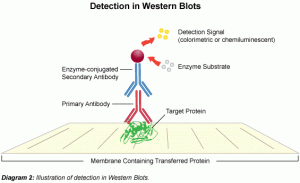
Western blotting uses antibodies to detect specific proteins separated by electrophoresis.
Procedure overview:
-
proteins are separated by SDS-PAGE
-
proteins are transferred to a membrane
-
primary antibodies bind to target proteins
-
enzyme-linked secondary antibodies generate a detection signal
This technique provides information about protein size and expression levels.
ELISA (Enzyme-Linked Immunosorbent Assay)
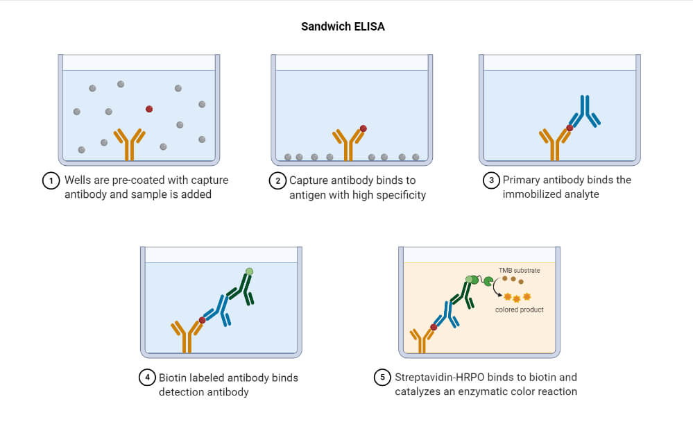
Antigen is immobilized on microplates; primary antibody binds the analyte; enzyme-linked secondary antibody catalyzes a colorimetric reaction proportional to analyte concentration. Sandwich, competitive, and indirect formats are widely used for quantification.
Choosing the Right Secondary Antibody
Selecting the correct secondary antibody is critical for experimental success.
Researchers should consider several factors:
Host Species
The secondary antibody must recognize the host species of the primary antibody.
Example:
-
rabbit primary antibody → anti-rabbit secondary antibody
Detection Label
Common secondary antibody labels include:
-
HRP (horseradish peroxidase)
-
alkaline phosphatase
-
fluorescent dyes (FITC, Alexa Fluor)
Cross-Reactivity
Secondary antibodies should be highly specific to avoid cross-reactivity with other immunoglobulins in the sample.
Best Practices for Antibody Experiments
To ensure accurate and reproducible results, laboratories should follow best practices when using antibody systems.
Key recommendations:
-
optimize antibody dilution concentrations
-
include positive and negative controls
-
use proper blocking buffers to reduce background
-
validate antibody specificity
-
store antibodies under recommended conditions
Proper experimental design significantly improves data reliability.
Conclusion
Primary and secondary antibodies form the backbone of many laboratory detection techniques. Their combined use enables highly sensitive and specific identification of proteins in complex biological samples.
By understanding the differences between these antibody types and applying best practices in antibody selection and optimization, researchers can achieve more reliable experimental results and deeper insights into biological processes.
Advances in antibody engineering and detection technologies continue to expand the possibilities of antibody-based research, making them indispensable tools in immunology, molecular biology, and biomedical diagnostics.