
Introduction
Autoimmune diseases arise when the immune system mistakenly targets the body’s own tissues. A key hallmark of many autoimmune disorders is the presence of autoantibodies, antibodies produced by the immune system that react against self-antigens. These antibodies can serve as important biomarkers for disease diagnosis, monitoring, and research.
In clinical laboratories and biomedical research, the detection and characterization of autoantibodies provide valuable insights into disease mechanisms and immune dysfunction. Techniques such as immunofluorescence assays, ELISA, and Western blotting allow scientists to identify and quantify specific autoantibodies associated with conditions such as type 1 diabetes, autoimmune gastritis, and endocrine disorders.
This article explores the biological mechanisms behind autoantibody formation, their role in autoimmune diseases, and the laboratory methods used to detect and analyze them.
Understanding Autoantibodies
What Are Autoantibodies?
Autoantibodies are immunoglobulins produced by B lymphocytes that recognize and bind to self-antigens, molecules naturally present in the body. Under normal physiological conditions, the immune system maintains tolerance toward self-components. However, when immune tolerance fails, autoreactive B cells may produce antibodies against the body’s own proteins, enzymes, or cellular structures.
These antibodies can target a wide range of biological molecules, including:
-
intracellular enzymes
-
cell surface receptors
-
nuclear components
-
tissue-specific proteins
Some autoantibodies contribute directly to tissue damage, while others serve primarily as diagnostic markers of autoimmune activity.

Mechanisms Behind Autoantibody Formation
The generation of autoantibodies typically results from a breakdown of immune tolerance mechanisms. Several biological processes may contribute to this phenomenon.
Loss of Immune Tolerance
During immune development, lymphocytes that strongly react with self-antigens are usually eliminated or inactivated. This process, known as central and peripheral tolerance, prevents autoimmune responses. When these mechanisms fail, autoreactive B cells may persist and produce autoantibodies.
Molecular Mimicry
Infections may trigger autoimmunity through molecular mimicry, a mechanism in which microbial antigens resemble host proteins. As a result, antibodies generated against pathogens may also recognize similar self-antigens.
Genetic Susceptibility
Certain genetic backgrounds increase susceptibility to autoimmune diseases. Variations in genes involved in immune regulation can predispose individuals to produce autoantibodies.

Common Autoantibodies Associated with Autoimmune Diseases
Different autoimmune diseases are associated with distinct autoantibody profiles. Identifying these antibodies helps clinicians and researchers better understand disease pathology.
Islet Cell Antibodies (ICA)
Islet cell antibodies target pancreatic beta cells responsible for insulin production. Their presence is strongly associated with type 1 diabetes and may appear years before clinical symptoms develop.
Glutamic Acid Decarboxylase Antibodies (GAD)
Autoantibodies against glutamic acid decarboxylase are also frequently detected in type 1 diabetes. They are commonly used as diagnostic markers in autoimmune diabetes research.
Gastric Parietal Cell Antibodies
These antibodies target gastric parietal cells, which produce stomach acid and intrinsic factor. They are often linked to autoimmune gastritis and pernicious anemia.
Adrenal Antibodies
Adrenal antibodies may attack the adrenal cortex and are associated with Addison’s disease, a disorder affecting hormone production.
Laboratory Methods for Detecting Autoantibodies
Modern immunology laboratories rely on several analytical techniques to identify and quantify autoantibodies.
Immunofluorescence Assays
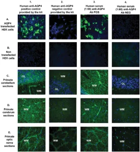
Indirect immunofluorescence is one of the most widely used techniques for detecting autoantibodies. In this method, patient serum is incubated with antigen-containing tissue sections or cultured cells. If autoantibodies are present, they bind to their target antigens.
A fluorescently labeled secondary antibody is then applied, allowing researchers to visualize antibody binding using fluorescence microscopy.
Advantages:
-
high sensitivity
-
visualization of antibody patterns
-
useful for screening multiple autoantibody types
Enzyme-Linked Immunosorbent Assay (ELISA)
ELISA is a quantitative method widely used for detecting specific antibodies in biological samples.
In this assay:
-
antigens are immobilized on a microplate
-
patient serum is added
-
antibodies bind to the antigen
-
enzyme-linked secondary antibodies produce a measurable color signal
ELISA offers several advantages:
-
high throughput analysis
-
quantitative measurement of antibody levels
-
strong reproducibility
Western Blot Analysis
Western blotting is often used as a confirmatory technique for detecting autoantibodies against specific proteins.
Proteins are first separated by electrophoresis and transferred to a membrane. Patient serum is then applied to identify antibodies that recognize specific protein bands.
This technique provides:
-
high specificity
-
confirmation of antigen identity
-
molecular weight information
Importance of Autoantibody Detection in Research and Diagnostics
Autoantibody analysis plays a crucial role in both clinical diagnostics and biomedical research.
Early Disease Detection
Many autoimmune diseases develop gradually, and autoantibodies may appear long before symptoms occur. Detecting these markers allows for earlier diagnosis and monitoring.
Disease Classification
Different autoantibody profiles help classify autoimmune disorders and distinguish between related conditions.
Therapeutic Research
Autoantibodies provide valuable information for developing targeted therapies and immunomodulatory treatments.
Best Practices for Autoantibody Research in the Laboratory
To obtain reliable results, researchers should follow several best practices when working with antibody-based assays.
Key recommendations:
-
validate antibodies before experimental use
-
include positive and negative controls
-
optimize antibody concentrations
-
use appropriate blocking reagents to minimize background signals
-
maintain proper storage conditions for antibody reagents
Careful experimental design ensures reproducibility and improves the reliability of scientific findings.
Conclusion
Autoantibodies play a central role in the development and diagnosis of autoimmune diseases. Understanding their mechanisms, targets, and detection methods enables researchers and clinicians to better characterize immune disorders and improve patient care.
Through advanced laboratory techniques such as immunofluorescence microscopy, ELISA assays, and Western blotting, scientists can accurately detect and study autoantibody activity. These analytical approaches continue to drive progress in immunology, autoimmune disease research, and biomarker discovery.
As scientific technologies evolve, the study of autoantibodies will remain a fundamental component of modern biomedical research.