Thyroid Antibodies (Thyroid Autoantibodies)
Introduction :
Thyroid antibodies are immunoglobulins produced by the immune system that target proteins within the thyroid gland. These antibodies are crucial biomarkers for diagnosing autoimmune thyroid diseases, such as Hashimoto’s thyroiditis, Graves’ disease, and postpartum thyroiditis. Immunofluorescence techniques allow researchers and clinicians to visualize these antibodies in tissue sections, providing insight into autoimmune mechanisms and enabling early detection of disease.
This page provides a detailed educational resource for laboratory scientists, medical professionals, and researchers, with images, videos, and practical tips for detecting and interpreting thyroid autoantibodies.

1. Overview of Thyroid Antibodies
Thyroid autoantibodies can be classified based on their target antigen:
| Antibody Type | Target | Associated Disease | Detection Method |
|---|---|---|---|
| Anti-Thyroglobulin (TgAb) | Thyroglobulin in follicular lumen | Hashimoto’s thyroiditis, Graves’ disease | IIF, ELISA, Western blot |
| Anti-Thyroid Peroxidase (TPOAb) | Thyroid peroxidase enzyme | Hashimoto’s thyroiditis | IIF, ELISA |
| TSH Receptor Antibodies (TRAb) | TSH receptor on follicular cells | Graves’ disease | IIF, ELISA, bioassay |
| Pendrin Antibodies | Pendrin protein in thyroid | Rare autoimmune thyroid disorders | IIF, Western blot |
| Sodium-Iodide Symporter Antibodies (NIS-Ab) | NIS protein on thyroid membrane | Experimental/Research | IIF, ELISA |
2. Anti-Thyroglobulin Antibodies (TgAb)
Function:
Thyroglobulin is a large glycoprotein stored in the thyroid follicle lumen, essential for thyroid hormone synthesis. TgAb binds to thyroglobulin and can contribute to thyroid tissue damage.
Clinical Significance:
-
Present in ~80% of patients with Hashimoto’s thyroiditis.
-
Can also be detected in some Graves’ disease patients.
-
High TgAb titers may indicate ongoing autoimmune activity or risk of hypothyroidism.
Immunofluorescence Pattern:
-
Granular cytoplasmic staining within thyroid follicles.
-
Colloid appears brightly fluorescent when bound by patient IgG.
3. Anti-Thyroid Peroxidase Antibodies (TPOAb)
Function:
TPO is an enzyme on the apical surface of thyroid follicular cells, involved in iodination of thyroglobulin and hormone synthesis. TPOAb interferes with this process, contributing to hypothyroidism.Clinical Significance:
-
Most prevalent antibody in Hashimoto’s thyroiditis.
-
Useful marker for predicting progression of autoimmune thyroid disease.
-
Detected in both subclinical and clinical thyroiditis.
Immunofluorescence Pattern:
-
Granular cytoplasmic fluorescence along follicular membranes.
-
Strong staining correlates with high TPOAb titer.

Expression levels of TPO in thyroid cancer and normal thyroid tissues. 4. TSH Receptor Antibodies (TRAb)
Function:
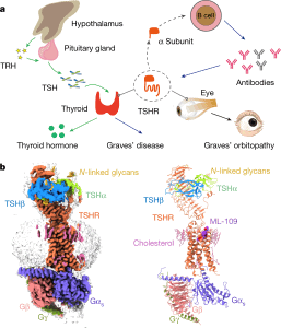
TRAb are immunoglobulins that bind to the TSH receptor on thyroid follicular cells, stimulating (or sometimes blocking) thyroid hormone production.Clinical Significance:
-
Primary marker for Graves’ disease.
-
Correlates with disease activity and severity.
-
Can be measured with ELISA, IIF, or bioassays.
Immunofluorescence Pattern:
-
Membrane-specific fluorescence around thyroid follicular cells.
-
-
Stimulating TRAb produces uniform bright staining on the membrane.
-
5. Advanced/Experimental Thyroid Antibodies
5.1 Pendrin Antibodies
-
Target the pendrin protein in the thyroid membrane.
-
Studied mostly in rare autoimmune thyroid conditions.
5.2 Sodium-Iodide Symporter Antibodies (NIS-Ab)
-
Target NIS protein, involved in iodide uptake.
-
Relevant in research on thyroid autoimmunity and radioiodine therapy.
6. Laboratory Techniques for Detection
6.1 Indirect Immunofluorescence (IIF)
Workflow:
-
Patient serum applied to thin thyroid tissue sections.
-
IgG antibodies bind target antigens.
-
Fluorescent secondary antibody applied.
-
Staining patterns visualized under a fluorescence microscope.
Tips:
-
Include positive and negative controls.
-
Use high-quality fluorophores to prevent fading.
-
Capture images with high-resolution camera and consistent exposure.
-
-
6.2 ELISA for Thyroid Antibodies
-
Quantitative method to measure TgAb, TPOAb, and TRAb.
-
High sensitivity and specificity.
Useful for longitudinal monitoring of autoimmune thyroid disease.
6.3 Western Blot
- Confirms specific antigen-antibody interaction.
- Helps distinguish cross-reactivity.
7. Clinical Relevance and Case Studies
7.1 Hashimoto’s Thyroiditis
-
Autoimmune destruction of thyroid follicles.
-
Presence of TgAb and TPOAb indicates active autoimmune process.
-
Early detection allows timely treatment with thyroid hormone replacement.
7.2 Graves’ Disease
-
Overproduction of thyroid hormone due to stimulating TRAb.
-
Immunofluorescence confirms TRAb activity and predicts disease course.
7.3 Postpartum Thyroiditis
-
Temporary thyroid dysfunction after childbirth.
-
TgAb and TPOAb may predict onset.
-
8. Tips for Laboratory Best Practices
-
Sample Handling: Preserve serum samples at -80°C to maintain antibody integrity.
-
Standardization: Use consistent tissue sections, incubation times, and antibody concentrations.
-
Documentation: Annotate images with antibody type, tissue type, and magnification.
-
Safety: Follow biosafety regulations for handling human samples.
-